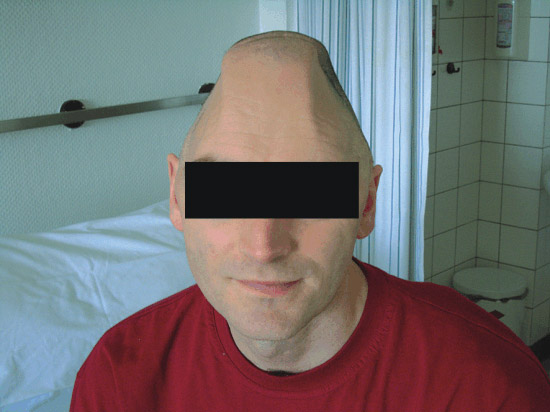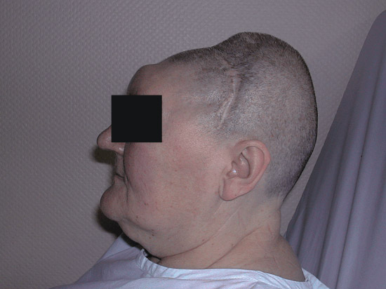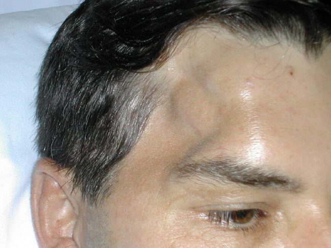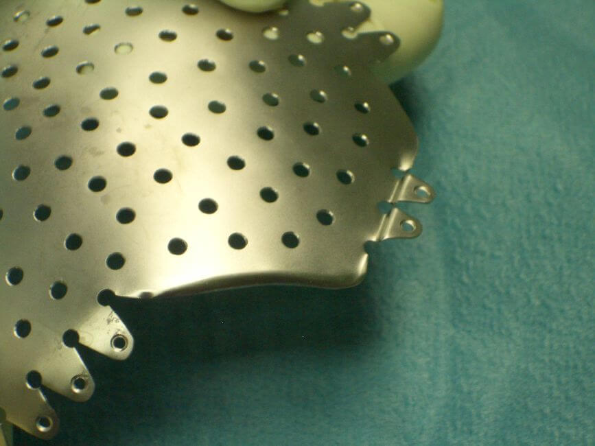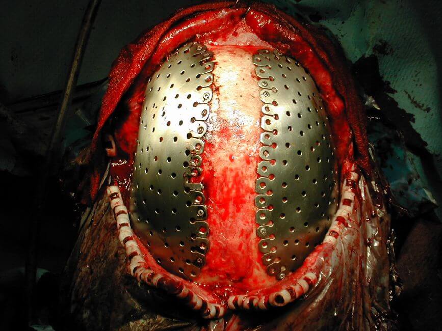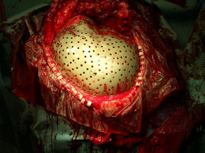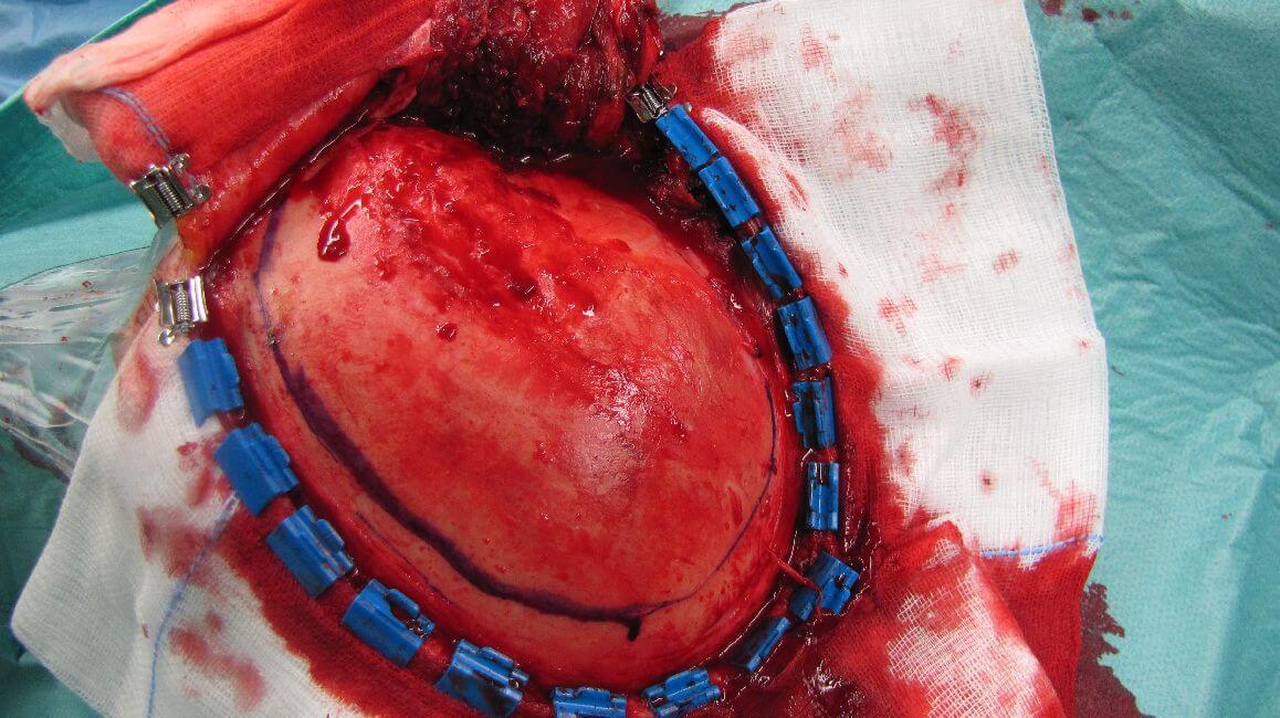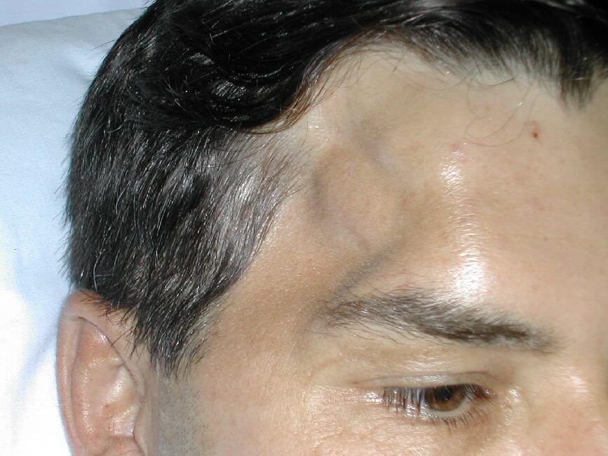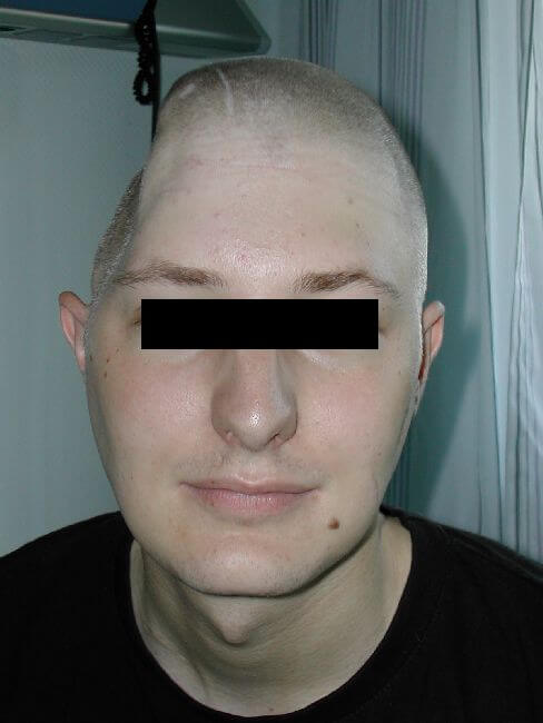
Defect of the cranial vault
Dolor anterdum, lacus et vulputate nulla commodo sem, at egestas nulla metus vel sapien.


Dolor anterdum, lacus et vulputate nulla commodo sem, at egestas nulla metus vel sapien.
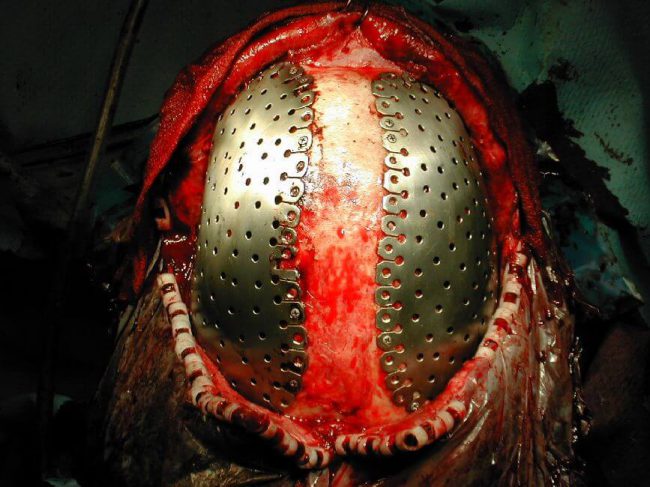
Case StudiesBilateral defect; reconstruction of temporal muscle After decompressive bilateral craniectomy because of generalized brain edema; infection of the reimplanted autologous skull flaps and their removal; massive subgaleal hygromas on both sides. In order to anticipate atrophy of the temporal muscles their reconstruction has to be planned during the manufacturing of cranioTOP® that means that…
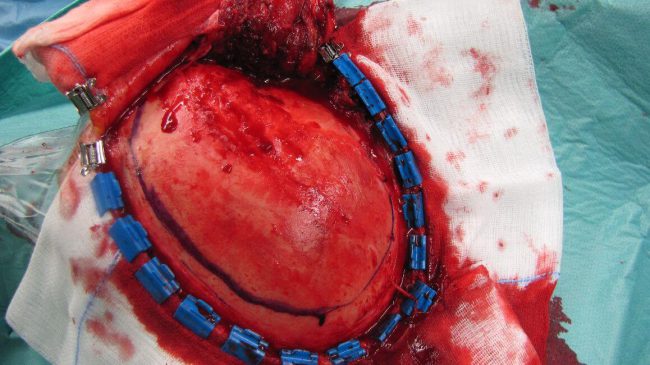
Case StudiesOsteolysis bilateral After decompressive craniectomy because of traumatic brain edema, osteolysis of the reimplanted autologous skull flaps. Neurosurgical Clinic of the City of Dortmund (Priv.-Doz.Dr.R.Schultheiß)
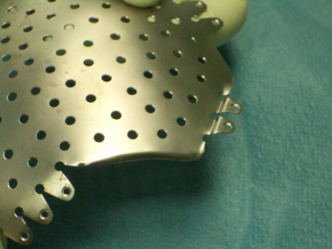
Case StudiesFixation of temporal muscle, dura and galea to cranioTOP® At the perforations of cranioTOP® the temporal muscle can be attached by sutures when no reconstruction is necessary or desired: with the same technique the dura and the galea can be attached in order to avoid subdural or subgaleal hygromas or hematomas. Neurosurgical Clinic of…
Case StudiescranioTOP® used as a template for resection of tumorous skull flap (template for craniotomy) In case of a mainly osseous sphenoid wing meningeoma partially with exophytic growth. Neurosurgical Clinic of the University Hospitals of Regensburg, (Dr.K.-M.Schebesch und Prof.Dr.A.Brawanski)
Case StudiesBilateral cranioplasty;worst-case scenario for CT-and MRI-compatibility Decompressive craniectomies in the course of generalised brain edema; months later reimplantation of the autologeous skull flaps , but removal nesessary because of wound infection. A „historical“ case occurring during the early developmental state of cranioTOP® ,when rather thick (0,7mm) sheets of titanium were used as half stuff(meanwhile…
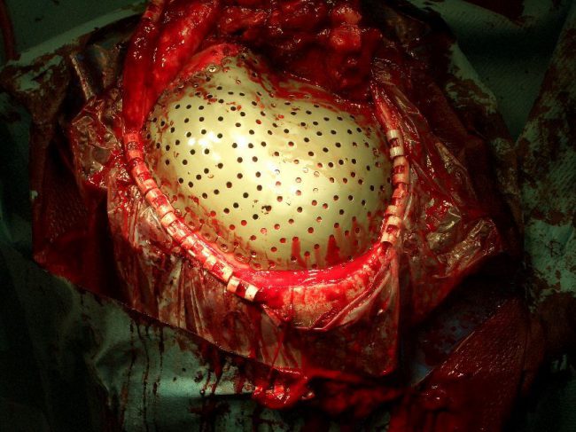
Case StudiesBifrontal cranioTOP®; MRI-Compatibility After 11 operations, at first total excision of a fronto-basal meningeoma including reimplantation of the autologeous skull flap; wound infection and removal of the flap, several trials with PMMA-cranioplasties every time with local infection and rejection with resulting removals again. Scar tissue contraction has to be anticipated while planning cranioTOP®, that…
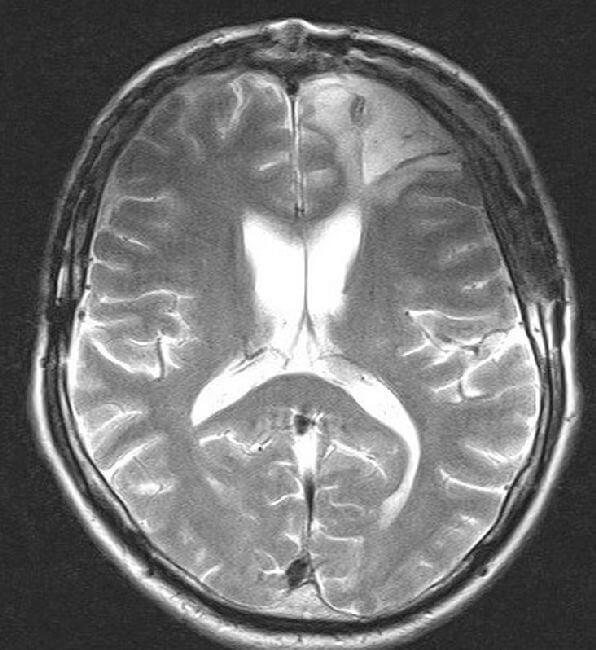
Case StudiesBilateral cranioTOP®; worst case scenario for CT-and MRI-Compatibility? Postoperative MRI documentation prooves the minor susceptibility to metal artifacts of even very extensive cranioTOP® cranioplasties; the following study was performed with PICKER Eclipse 1,5 Tesla; in all relevant sequences and slices no significant impairment of the image quality was found. Neurosurgical Clinic of the City…
Case StudiesPterional defect After subarachnoid hemorrhage, clipping of an aneurysm, infection and removal of the bone flap, insufficient trial of cranioplasty with ceramic material which has fractured and sunk and is still in place. Neurosurgical Clinic of the City of Dortmund (Priv.-Doz.Dr.R.Schultheiß)